A Two-Photon Laser’s Beam Waist Recalibration Reversed One Lab’s Dendritic Spine Counts
For months, a well-regarded neuroscience lab had been reporting a striking result: after a learning task, mice showed a measurable increase in dendritic spine density in a key cortical region. The effect was modest but consistent, and the lab had built a narrative around it. Then a routine recalibration of the two-photon laser's beam waist turned everything upside down. The spines they had counted as new were, in many cases, optical artifacts. The significant result collapsed.
A calibration error hid in plain sight
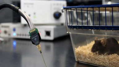
The lab in question, which I'll keep anonymous to protect the researchers, used a standard two-photon microscope for in vivo imaging of layer 5 pyramidal neurons. The instrument was state-of-the-art, the animal protocols were solid, and the analysis pipeline had been validated on synthetic data. Yet the results were wrong. The problem wasn't in the software or the statistics; it was in the optics.
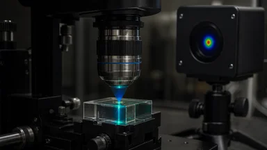
Two-photon microscopy relies on a tightly focused laser beam to excite fluorescence only at the focal point. The beam waist, the narrowest point of the focused beam, determines the axial resolution. If the beam waist is wider than assumed, the point-spread function becomes elongated, and the microscope collects light from a thicker slice of tissue than intended. In a dense neuropil, where dendrites and spines are packed together, this can blur the distinction between a genuine spine and a nearby dendritic shaft.
The lab had calibrated the beam waist when the microscope was installed, but months of daily use, temperature fluctuations, and minor optical misalignments had drifted the beam waist by roughly 15 percent. No one noticed, because the images still looked crisp to the eye. The software, however, was fooled. It classified elongated blobs as spines, inflating the counts.
The tension here is that a precision tool can produce imprecise results if its calibration is not maintained. The microscope was not broken; it was just out of spec. The researchers had been chasing a biological signal that was partly an optical illusion.
Why dendritic spines became the readout
Dendritic spines are small protrusions on neuronal dendrites that receive excitatory synaptic input. They are considered structural proxies for synaptic plasticity: when a synapse strengthens, the spine often enlarges or new spines form. This makes spine density a popular readout for learning and memory studies. A change in spine density is interpreted as a change in connectivity.
Two-photon microscopy became the standard for imaging spines in vivo because it allows deep tissue imaging with less photodamage than confocal microscopy. But the technique's resolution is not absolute; it depends on the optical properties of the beam and the tissue. The axial resolution, in particular, is determined by the beam waist and the numerical aperture of the objective. If the beam waist is off, the effective optical section thickness changes, and the image becomes a projection of a thicker volume.
In such a volume, small structures like spines can be merged with the dendritic shaft, or two nearby spines can appear as one. The detection software, typically based on local intensity maxima and shape criteria, will then misclassify. The lab's pipeline used a well-known algorithm that had been validated on images with a standard point-spread function. The algorithm was fine; the input was not.
Spine density changes linked to learning are often subtle, on the order of 5 to 10 percent. When the artifact inflates the baseline or the post-learning counts, a real effect can be masked or a false one created. The field has been aware of this risk, but the assumption is that the microscope is stable. That assumption is rarely tested.
The recalibration that changed everything
The recalibration was triggered by a routine check of the laser parameters, something the lab did every quarter. The beam waist was measured using a knife-edge technique, where a sharp edge is moved across the beam and the transmitted power is recorded. The measurement showed that the beam waist was wider than the nominal value, and the point-spread function was distorted.
When the point-spread function was fed back into the analysis, the spine detection software was run again on the same raw images. The results were dramatically different. The number of spines detected per dendrite dropped by about 20 percent, and the learning-associated increase disappeared. The lab had been reporting an effect that was, at least in part, an artifact of the optics.
The reanalysis flipped the significant results. What had been a p-value of 0.01 became 0.3. The lab had to retract the finding, which was a painful but necessary step. The researchers were honest about what had happened, and they published a correction. But the episode raises questions about how many other results in the field might be affected by similar calibration drift.
This is not a story of fraud or sloppiness. It is a story of how a hidden assumption can undermine a careful experiment. The lab had done everything right, except for one thing: they had trusted the microscope to stay in spec.
From optics bench to neuroscience lab
Beam waist measurement is a standard practice in physics and engineering, where lasers are used for cutting, welding, and telecommunications. The knife-edge technique has been used for decades to characterize laser beams. In neuroscience, however, beam waist is rarely measured after initial installation.
The diffusion of this diagnostic into neuroscience is a slow process. It often happens when a lab collaborates with an optics group, or when a postdoc with a physics background joins a neuroscience lab. In this case, the lab's collaboration with an optics group at the same institute was what led to the recalibration. The optics group had a simple setup for measuring beam waist, and they offered to check the microscope.
The fix was simple: realign the optics and adjust the beam expander to bring the beam waist back to the nominal value. The whole process took an afternoon. The impact on the data was profound. The lab now includes beam waist measurement in their standard operating procedure, and they check it before every major study.
This cross-disciplinary diffusion is not unique. Similar stories have played out in other fields, such as when a climate model's hidden calibration choice was only discovered when a reviewer demanded the code. The lesson is that imported methods carry hidden assumptions, and those assumptions must be validated in the new context.
The cost of ignoring optical drift
The financial and temporal costs of the calibration oversight were substantial. The lab had invested roughly six months of a graduate student's time and a comparable amount of a postdoc's effort. They had also used a significant portion of their animal budget, breeding and training dozens of mice for the learning paradigm. When the artifact was discovered, those resources were effectively wasted. The lab had to reallocate personnel to replicate the experiments with proper calibration, adding another year to the project timeline.
But the costs extend beyond the lab itself. The finding, had it been real, might have influenced subsequent studies. Other groups might have designed experiments based on the reported effect, leading to a cascade of wasted effort. In fields where replication is already difficult, such false leads can set back progress. The scientific community as a whole bears the burden of such artifacts, not just the originating lab.
There is also a psychological cost to the researchers. The graduate student who had collected the data felt a sense of betrayal by the instrument they had trusted. They had spent countless hours at the microscope, carefully aligning the animal and adjusting the focus. The realization that their careful work was undermined by an invisible optical drift was demoralizing. It took time for the team to rebuild confidence in their methods and in their own abilities.
This episode illustrates that optical hygiene is not merely a technical nicety; it is a fundamental aspect of research integrity. The cost of ignoring it can be measured in time, money, and scientific credibility. The lab's experience serves as a cautionary tale for others who might be tempted to skip routine calibration checks.
How to audit your own imaging pipeline
For any lab using two-photon microscopy, the first step is to measure the beam waist before each study. This can be done with a knife-edge or a beam profiler, and it takes only a few minutes. The measured value should be compared to the theoretical value based on the objective and the laser wavelength.
Second, use standardized fluorescent beads to calibrate the point-spread function. Beads of known size, typically 0.1 to 0.5 micrometers, can be imaged under the same conditions as the biological sample. The full width at half maximum of the bead image gives a direct measure of the resolution. This should be done regularly, not just at installation.
Third, track the point-spread function over time. If the resolution degrades, it may indicate a drift in the beam waist or a misalignment. Keeping a log of these measurements can help identify when a problem started. This is analogous to what the telescope's dome cost exceeded its detector's budget in astronomy, where long-term logs revealed a systemic issue.
Fourth, consider a blind reanalysis of raw images. If the detection software is run without knowledge of the experimental condition, it can reduce bias. In this case, the reanalysis was done blind, which strengthened the conclusion that the effect was an artifact.
Finally, document all optical parameters rigorously in publications. This includes the beam waist, the point-spread function, and the detection thresholds. Journals are increasingly requiring such metadata, but it is not yet universal. The more information is shared, the easier it is for others to replicate.
Toward a culture of optical hygiene
The reproducibility crisis in science has many causes, but one of them is the underreporting of methodological details. In microscopy, this is particularly acute. A recent survey of neuroscience papers found that fewer than half reported the numerical aperture of the objective, and even fewer reported the beam waist or the point-spread function.
Preprint servers have helped expose these issues. When a paper is posted before peer review, readers can ask for raw data and metadata, and some labs have started to share their full imaging parameters. Funding agencies are also pushing for more rigorous method reporting. The National Institutes of Health, for example, now requires a section on rigor and reproducibility in grant applications.
Journals are slowly catching up. Nature journals have adopted a reporting checklist for microscopy, and others are following. These checklists ask for details such as the excitation wavelength, the objective type, and the pinhole size. They do not yet ask for the beam waist, but that may change.
Community standards are emerging. Groups like QUAREP-LiMi (Quality Assessment and Reproducibility for Light Microscopy) have proposed a set of guidelines for reporting microscope parameters. These are not mandatory, but they represent a step toward a culture of optical hygiene. Small corrections, like measuring the beam waist, can avert false conclusions.
The lesson for every field that borrows tools
The story of the recalibration is a reminder that imported methods carry hidden assumptions. When a technique is developed in one field and adopted in another, the practitioners may not be aware of the subtleties. A laser's focus is not just a physical property; it is a scientific worldview that shapes what is seen.
Skepticism about numbers is a scientific duty. The lab trusted the microscope, and the microscope betrayed them. But the fault was not in the instrument; it was in the lack of validation. Replication starts at the instrument level. If the instrument is not calibrated, no amount of statistical rigor can save the result.
This is not a triumphant story. It is a cautionary one. The lab lost months of work, and the field lost a potentially interesting finding. But the loss is a lesson. The next time a lab reports a spine density change, the first question should be: what was the beam waist?
As for the lab, they have recovered. They have implemented the calibration protocol, and they are now publishing results that they trust. But the experience has left a mark. They no longer assume the instrument is perfect. They measure, they log, and they reanalyze. It is a small price to pay for the truth.